Transient Osteoporosis Of The Hip Mri

Transient Osteoporosis Of The Hip Radiology Reference Article Radiopaedia Org
Transient Osteoporosis Of The Hip Orthoinfo Aaos
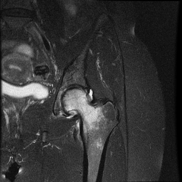
Idiopathic Transient Osteoporosis Of The Hip Radiology Case Radiopaedia Org
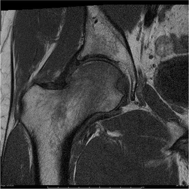
Transient Osteoporosis Of The Hip Underlying Subchondral Fracture Radiology Case Radiopaedia Org

Transient Osteoporosis Not Just The Hip To Worry About Sciencedirect
Transient Osteoporosis Of The Hip Image Radiopaedia Org
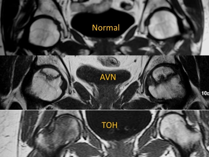
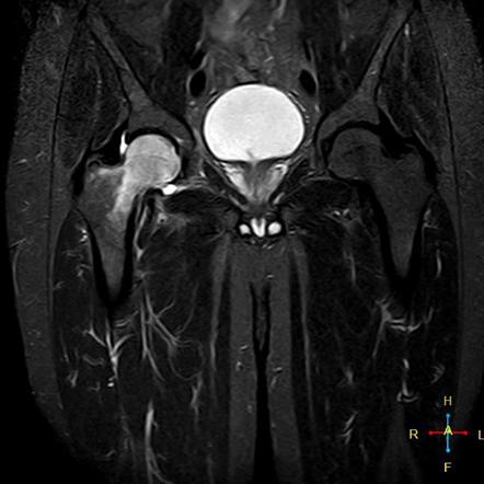
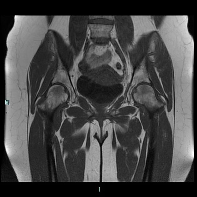
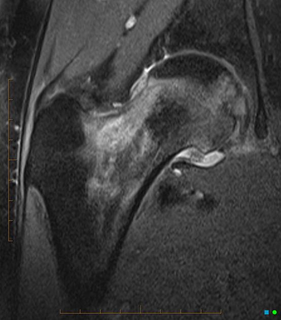
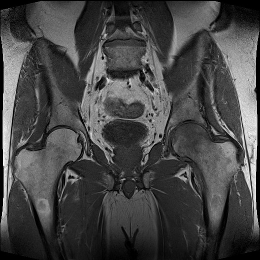
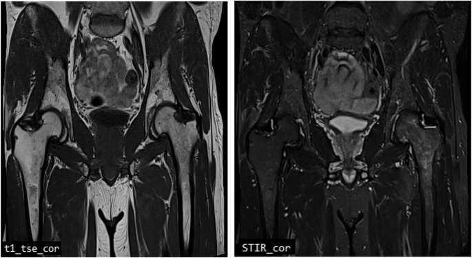
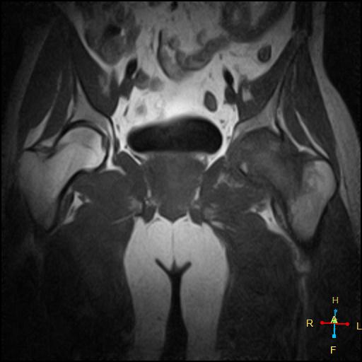
Because of this mri is one of the most useful studies to help diagnose the condition.
Transient osteoporosis of the hip mri.
Http Canjsurg Ca Wp Content Uploads 2014 03 46 3 187 Pdf
Idiopathic Transient Osteoporosis Of The Hip Radiology Case Radiopaedia Org
Https Www Journalofosteopathicmedicine Com Article S1746 0689 15 00133 9 Pdf
Http Www Akot Com Ar Cokiba Cursos 2018 16 Curso Oyt Files Transient 20osteoporosis 20of 20the 20hip 20review 20of 20the 20literature Pdf

Pdf Transient Osteoporosis Of The Hip
Transient Osteoporosis Of Hip

Annals Of Rehabilitation Medicine
Transient Hip Osteoporosis Radiology Case Radiopaedia Org

Transient Osteoporosis Of Pregnancy In A 34 Year Old Female Sciencedirect
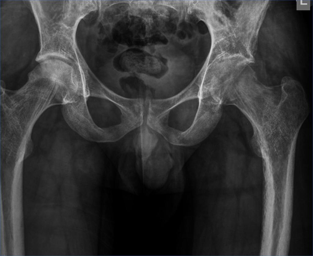
Transient Osteoporosis Of The Hip Radiology Case Radiopaedia Org
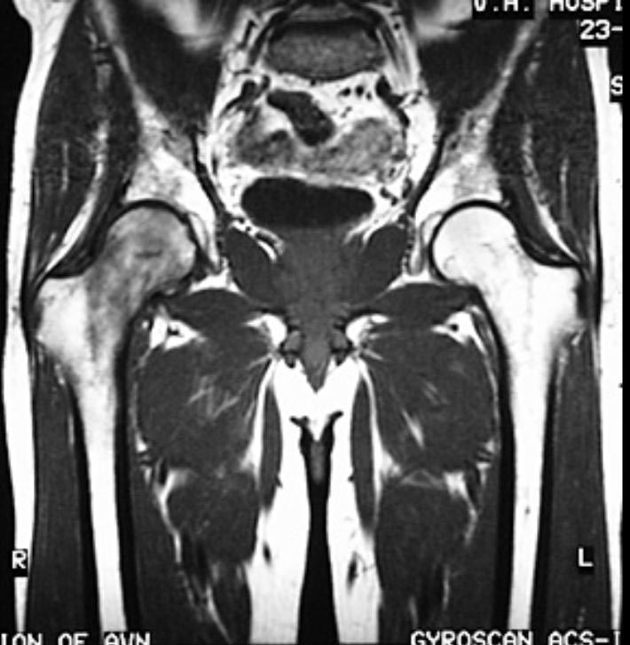
Bilateral Transient Osteoporosis Of The Hip Postpartum Radiology Case Radiopaedia Org

Full Text Transient Osteoporosis Of The Hip Risk And Therapy Oarrr
Transient Migratory Osteoporosis Of The Hip And Talus A Case Report Journal Of Orthopaedic Case Reports
Transient Bone Marrow Edema Of The Hip Radiology Case Radiopaedia Org

File Stir Mri Of Transient Osteoporosis Jpg Wikipedia

Idiopathic Transient Osteoporosis Of The Hip Post Partum Radiology Case Radiopaedia Org
Transient Idiopathic Osteoporosis Of The Hip Robert Howells

Transient Osteoporosis Of The Hip After Bariatric Surgery Sciencedirect
Https Encrypted Tbn0 Gstatic Com Images Q Tbn 3aand9gcqgyzo8ptjslcsutaf29vh9e24aaqcbd 39zdpzsmxvumkuzf5n Usqp Cau

Pdf Evaluation Of Transient Osteoporosis Of The Hip In Magnetic Resonance Imaging Semantic Scholar

Imaging Of The Hip And Pelvis Radiology Key
Transient Osteoporosis Of The Hip And Subclinical Hypothyroidism An Unusual Dangerous Duet Case Report And Pathogenetic Hypothesis Bmc Musculoskeletal Disorders Full Text
Idiopathic Transient Osteoporosis Of The Hip Image Radiopaedia Org

Arm Annals Of Rehabilitation Medicine
Source : pinterest.com
